3D Cone Beam and 3D Dental Scans Santa Rosa, CA
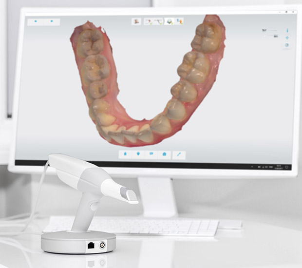
Advancements in dental technology have brought about many new tools that make it easier for dentists and more comfortable for patients. Dental cone beam computed tomography (CBCT), a new type of X-ray equipment, allows dentists to see a clear, detailed, three-dimensional image of the mouth without causing pain to the patient. This 3D imaging system takes full photos of the teeth, soft tissues, nerve pathways, and bones in a single scan.
Cone beam dental scans are available at Sai Dental Care in Santa Rosa and the surrounding area. Our staff can help you learn more about the procedure you are undergoing and answer any questions you have about 3D imaging. Call us at (707) 509-1146 to schedule a consultation appointment today.
How 3D Imaging Works
A 3D cone beam machine resembles conventional CT scan machines and comes in two different structures: an upright chair for sitting or a moveable table for lying down. Depending on the procedure and type of machine being used, the patient will be seated in an exam chair or lie down on an exam table. The chair has an extendable arm (C-arm) while the table has a rotator (gantry) that both rotate 360 degrees around the patient’s head, taking multiple images at once.
The images are taken at different angles and gathered to create a single 3D image. According to the Radiology organization, "In a single rotation, the detector can generate anywhere between 150 to 200 high resolution two-dimensional (2-D) images, which are then digitally combined to form a 3-D image that can provide your dentist or oral surgeon with valuable information about your oral and craniofacial health." The 3D image is available as soon as the scan is complete, allowing the doctor to discuss the patient’s treatment plan in the same visit.
“The chair has an extendable arm (C-arm) while the table has a rotator (gantry) that both rotate 360 degrees around the patient’s head, taking multiple images at once.”
Differences Between Traditional and Cone Beam
Traditional CT scanners and cone beam CT scanners both undertake the same basic function, but technical differences set them apart.
Traditional CT Scans
A traditional CT scan takes several pictures of internal body structures from multiple X-ray images generated by a computer. Atlantis, a radiology equipment company, explains, "The x-rays utilize radiation from a radioactive contrast injected into the body to create cross-sectional images." Traditional CT scans can offer a variety of benefits, primarily for surgeries and diagnostics.
Cone Beam CT Scans
Cone beam scanners use a cone-shaped beam radiating from an X-ray source, covering a wide range with just a single rotation around the patient’s head. The X-rays are compiled using a series of algorithms to furnish high-resolution 3D images. Cone beams use a fan beam, as opposed to a light beam, and emit 200-300 times less radiation.
“The X-rays are compiled using a series of algorithms to furnish high-resolution 3D images.”
What a Cone Beam Image Reveals
Cone beam CT scanners reveal far more information than traditional scanners and provide detailed images of a patient’s underlying bone structure. Cone beams are primarily used for cases in which traditional X-rays would not provide sufficient information needed for treatments, specifically surgeries and underlying disease. They can evaluate diseases of the jaw, dentition, body structures of the face, nasal cavity, and sinuses.
Cone beam technology has also been useful for diagnosing oral cancers and cysts and managing impacted teeth. A study on cone beam technology found that there has been a "significant contribution of CBCT in the planning and successful surgical management of dentigerous cysts and associated impacted teeth." Since cone beams take wide range photos, they capture in-depth areas that traditional scanners can easily miss.
“They can evaluate diseases of the jaw, dentition, body structures of the face, nasal cavity, and sinuses.”
Check out what others are saying about our dental services on Yelp: 3D Cone Beam and 3D Dental Scans in Santa Rosa, CA
What to Expect Post-Exam
The cone beam scan is painless and does not require anesthesia, allowing patients to resume normal activity right after their examination. Although the machine does not cause any pain, claustrophobic should inform their dental practitioner to better accommodate them.
After the exam, the dentist will write a report of the scan results. We will discuss the curated treatment plan with the patient, answering any questions they may have. The patient will be able to see their 3D images and follow the dentist as they move through the treatment plan, pointing at the treatment areas. Preferences for treatments, anesthetics, and medications will also be discussed to fast-track future visits.
“The patient will be able to see their 3D images and follow the dentist as they move through the treatment plan, pointing at the treatment areas.”
Questions Answered on This Page
Q. How does a 3D cone beam work?
Q. What is the difference between traditional and cone beam scanners?
Q. What can a cone beam scan reveal about a patient?
People Also Ask
Q. What does the dentist look for in a dental examination?
Frequently Asked Questions About 3D Cone Beam Scans
Q. Are cone beam scans covered by insurance?
A. Cone beam scans are increasingly being covered by insurance companies as there is a rise in the use of these scans with daily patients. However, not all insurance providers cover these types of scans. We recommend contacting your insurance provider to understand the details of your plan or request coverage for a cone beam scan.
Q. What kind of image does a cone beam scanner produce?
A. Cone beam scanners not only take images of the mouth but also the entire head. The positioning of your teeth affects the entire head, allowing dentists to see the underlying bone structure and jawline and creating more accurate treatments. They are especially useful for procedures around delicate areas and for planning correctional procedures.
Q. How long have cone beams been used?
A. Cone beam scanners are fairly new and not all dental clinics use them. Although they were invented in 1967, they were first introduced in the European market in 1996 and in the U.S. market in 2001. Today, about 72% of dentists use CBCT scanners.
Q. How safe are CBCT scans?
A. CBCT scans emit 200-300 times less or 1/10 the amount of radiation as traditional imaging systems and X-rays. Cone beam CT scanners also allow dentists to emit a certain dosage, making them safer and much more flexible. However, they still emit a certain amount of radiation and can be considered not safe, especially for children, pregnant women, and the elderly. Regardless, all X-ray scans in dentistry require some amount of radiation, and CBCT scans emit the lowest.
Q. Is there anything I should prepare for or bring for a CBCT exam?
A. There is no preparation required before a CBCT scan. It is important to remove all loose or metallic objects before the exam, as with any other X-ray exam. Patients with metal implants can get a CBCT scan. The dentist will ask you to stay still and do not swallow or talk during the exam.
Quality Dental Services Can Transform Your Smile
By visiting us as soon as possible, our team can help get you the professional treatment you need. Instead of waiting around and allowing the symptoms to get worse, we can provide you with treatment options.

Scan here to view this page, 3D Cone Beam and 3D Dental Scans, on mobile
Dental Terminology
Learn More About 3D Scans Today
If you are looking for more information pertaining to complete health dentistry or the use of 3D cone beams or 3D dental scans, call us at 707-509-1146.
Helpful Related Links
- American Dental Association (ADA). Glossary of Dental Clinical Terms. 2022
About our business and website security
- Sai Dental Care was established in 2013.
- We accept the following payment methods: American Express, Cash, Check, Discover, MasterCard, and Visa
- We serve patients from the following counties: Sonoma County
- We serve patients from the following cities: Santa Rosa, Fulton, Sebastopol, Cotati, and Cunningham
- Norton Safe Web. View Details
- Trend Micro Site Safety Center. View Details

Scan here to open directions to Sai Dental Care on mobile
Related Posts
How A Deep Dental Cleaning Is Performed
A deep dental cleaning is a way that dentists treat severe cases of gum disease. If your dentist detects signs of periodontitis, such as deep gum pockets and bone loss in the jaw, then they may recommend a cleaning to get the disease under control and so the patient can take the first step in…
The Importance Of Patient Compliance During Invisalign Treatment
Successful Invisalign treatment relies on daily habits, not just office visits. When a general dentist uses Invisalign to move teeth with clear aligners, patients need to follow instructions closely to keep the process on track. When patients wear aligners as directed and care for them properly, each stage of treatment can build on the previous…

Knocked Out Tooth Care From An Emergency Dentist
An emergency dentist can provide urgent care when a tooth is knocked out during sports, a fall, or another sudden accident. However, to increase the chances of saving the tooth, patients and dentists must act quickly. Knowing what to do before arriving at the dental office can help protect the root surface and improve the…
Explore additional topics covered on our website:
Call Us Today

Scan here to call Sai Dental Care on mobile